


Επιστροφή


Επιστροφή
The acromioclavicular (AC) joint is a small but important synovial joint located at the top of the shoulder. It is formed by the articulation between the acromion (the top part of the shoulder blade) and the distal (lateral) end of the clavicle. Like most joints in the human body, the AC joint is enclosed by a thin fibrous joint capsule. This capsule helps contain the synovial fluid, which lubricates the joint and facilitates smooth movement. Several ligaments stabilize the AC joint and help prevent dislocation:
Within the joint space lies a fibrocartilaginous disc, also referred to as the articular meniscus.
This disc varies in size and shape among individuals and acts as a shock absorber between the two bones, enhancing joint congruency and aiding in force transmission across the joint.
The acromioclavicular (AC) joint plays a crucial role in shoulder movement and stability, especially when lifting the arm or reaching overhead.
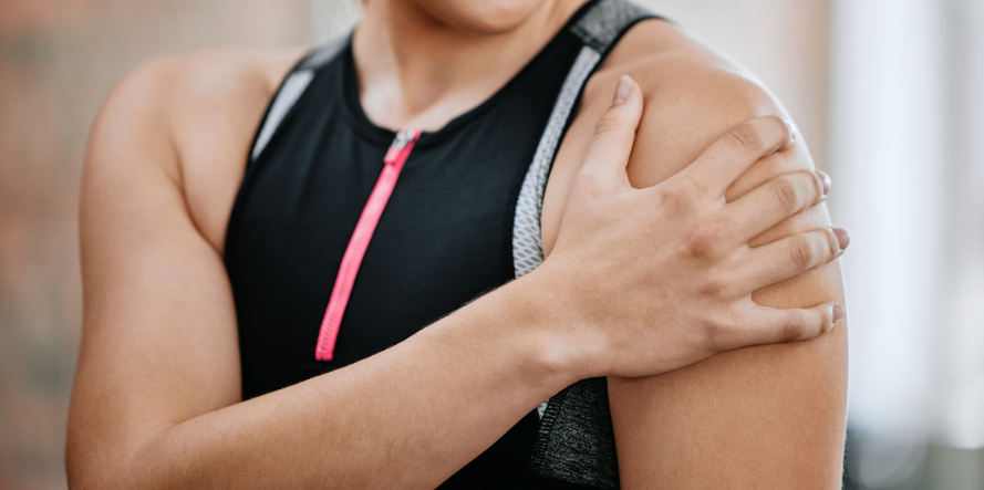
The most common cause of injury to the AC joint is a direct impact to the top of the shoulder, often due to a fall. This typically occurs when someone falls with the arm at their side, causing the force to travel through the shoulder and impact the joint directly. Sports like football, hockey, cycling, and skiing often see a higher incidence of AC joint injuries due to the nature of collisions or falls involved.
Mechanism of AC joint injury
This typically occurs when someone falls with the arm at their side, causing the force to travel through the shoulder and impact the joint directly. Sports like football, hockey, cycling, and skiing often see a higher incidence of AC joint injuries due to the nature of collisions or falls involved.
The severity of an AC joint injury depends on the extent of damage to the ligaments that stabilize the joint:
According to Rockwood’s Classification, there are six types of AC joint dislocations.
Your doctor will review your symptoms and medical history and perform a thorough physical examination to check for range of motion, stability, and strength of the joint. Initially, during examination, the characteristic prominence of the clavicle beneath the skin is often observed.
Palpation of the area is particularly painful. If necessary, your doctor will order certain imaging tests such as X-ray, MRI, CT scan, or ultrasound for a detailed evaluation of the joint and surrounding soft tissue structures to confirm the diagnosis. A radiograph of the contralateral (uninjured) shoulder is often taken for comparison.
The treatment of Acromioclavicular (AC) Joint Dislocation depends on the severity of the injury:
Physical therapy is important after immobilization or surgery to restore strength and range of motion. Most mild injuries heal well without surgery. Surgery in severe cases helps prevent chronic pain, weakness, and deformity. If untreated or improperly managed, chronic pain, arthritis, or shoulder dysfunction may develop.
After surgery, the arm is immobilized in a sling. This helps reduce pain, supports the joint, and protects the surgical repair by preventing movement that could disrupt healing.
From the second day, rehabilitation begins with physiotherapy and passive mobilization. This helps maintain joint flexibility and prevents stiffness without stressing the repaired tissues.
From the 2nd week, active strengthening exercises begin to rebuild the muscles around the shoulder and support joint stability. These exercises focus on the deltoid, trapezius, and rotator cuff muscles, which are critical for shoulder function.
Lifting light objects is usually allowed from the third week post-surgery, depending on healing progress. This gradual reintroduction helps the patient regain functional use without risking damage.
Full return to sports or high-demand activities generally happens after 6 to 8 weeks, when the joint is considered sufficiently healed and muscles are stronger.

Fill in your details below and we will contact you immediately!
WIN THE
MATCH POINT IN THE RACE
OF YOUR HEALTH!