


Επιστροφή


Επιστροφή
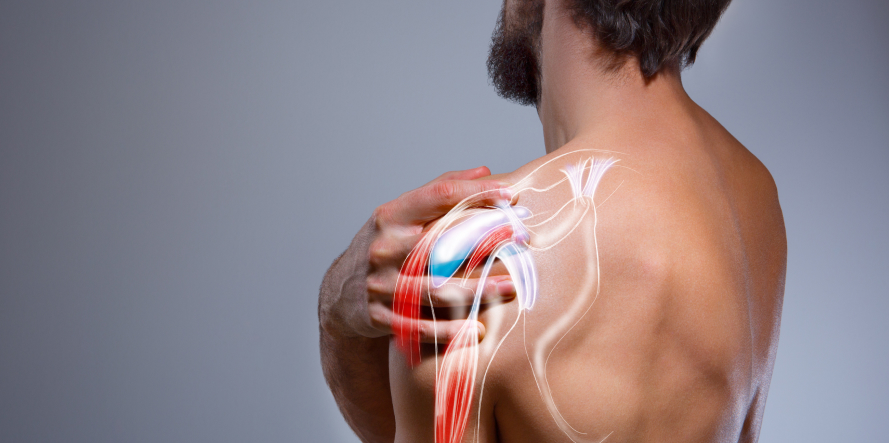
Shoulder Impingement Syndrome (SIS) is a condition where soft tissues in the shoulder joint become compressed or “impinged” between the bones during arm movement, leading to inflammation, pain, and restricted motion. This syndrome that occurs in the subacromial space caused by irritation of the rotator cuff tendons due to their “impingement” with the acromion.
The subacromial space is a narrow area located between the head of the humerus (upper arm bone) and the coracoacromial arch.
The coracoacromial arch includes the acromion (part of the scapula that extends over the shoulder joint), the coracoacromial ligament and the coracoid process (a bony projection from the scapula).
The inner subacromial space consists of critical structures such as rotator cuff tendons (especially the supraspinatus tendon), long head of the biceps tendon, subacromial bursa (a fluid-filled sac that reduces friction) and the coracoacromial ligament. These structures are surrounded by the subacromial bursa, which reduces friction between them.
Shoulder Impingement Syndrome (SIS) typically affects people between 30 to 50 years old as tendons become less elastic and more prone to injury with age. It also appears in people who engage in intense physical activity involving repetitive, high-intensity shoulder movements or work in manual labor jobs, where shoulder movements are continuous and strenuous.
It’s one of the most common causes of shoulder pain, accounting for roughly 60% of all shoulder-related complaints.
Shoulder Impingement Syndrome is caused by various factors that are divided into intrinsic (internal) and extrinsic (external) ones. People involved in overhead sports (e.g., swimming, tennis, baseball), manual labor (e.g., construction, painting) or repetitive shoulder activities are at higher risk due to frequent shoulder elevation and rotation, increasing the chance of tendon and bursa irritation.
More specifically, the causes include:
These originate within the shoulder joint and soft tissues:
These arise from structures outside the rotator cuff that contribute to compression:
The main cause of the syndrome is the narrowing of the space (<7 mm) between the head of the humerus and the acromion, which leads to inflammation of the subacromial bursa and mechanical irritation of the rotator cuff tendons, which pass through this space. Pain mainly occurs during abduction (arm lifting sideways) and forward flexion (lifting arm forward) of the upper limb, due to impingement of the humeral head against the anterior / inferior surface of the acromion.
The shoulder joint (glenohumeral joint) is the most mobile but least stable joint in the human body. Unlike other joints, its stability is ensured primarily not by the bones, but by the ligaments and muscles of the surrounding area.
Four muscles with their tendons (supraspinatus, infraspinatus, subscapularis and teres minor) form the rotator cuff muscles. In combination with the strong ligaments of the area, they stabilize the large-diameter head of the humerus within the small articular surface of the glenoid cavity. Due to this complex structure within a very limited space, the shoulder joint is susceptible to conditions such as Subacromial Impingement Syndrome (SAIS) or dislocation.
If not treated promptly and correctly, Shoulder Impingement Syndrome can lead to various complications such as:
The condition typically develops gradually and may be related to repetitive shoulder activity (common in athletes, manual laborers, or with aging). Here’s a breakdown of the listed symptoms:
If left untreated, Shoulder Impingement Syndrome (SIS) can lead to:
If you notice any of the above symptoms, it is essential to visit a specialized orthopedic surgeon for a proper diagnosis and treatment.
Shoulder Impingement Syndrome is a common cause of shoulder pain, particularly among athletes and individuals performing repetitive overhead activities. The diagnosis is multifaceted and involves a clinical evaluation, imaging tests, and sometimes additional diagnostic procedures to confirm the condition and rule out other shoulder pathologies.
The diagnostic process typically begins with a thorough clinical examination conducted by an orthopedic surgeon or a shoulder specialist. This step includes:
These tests aim to provoke pain by compressing the rotator cuff tendons under the acromion to confirm impingement.
Imaging is used to confirm clinical findings and assess the extent of structural abnormalities.
Shoulder X-rays are often the first imaging study ordered. They can reveal:
X-rays are useful for detecting bony changes, but they do not show soft tissue damage.
MRI provides a detailed view of the soft tissues and is usually performed if symptoms persist or if rotator cuff tear or bursitis is suspected. It helps in identifying:
MRI is particularly valuable for pre-surgical planning if conservative treatment fails.
The initial treatment of shoulder impingement syndrome (SIS) is usually conservative and involves a combination of non-surgical interventions aimed at relieving pain, reducing inflammation, and improving shoulder function.
The orthopedic surgeon may recommend temporarily avoiding activities that exacerbate symptoms and modifying daily activities to reduce stress on the shoulder. He may also prescribe non-steroidal anti-inflammatory drugs (NSAIDs) to manage pain and reduce inflammation. In addition, the patient follows a structured physical therapy program to strengthen the muscles around the shoulder, improve shoulder mechanics, and restore range of motion. Physical therapy may include stretching exercises, strengthening exercises, and techniques to improve posture and shoulder mechanics.
If symptoms persist despite initial treatments, a local injection of biological agents (platelet-rich plasma or PRP) may be used to further relieve persistent symptoms, reduce inflammation and promote healing.
Initially, Shoulder Impingement Syndrome (SIS) is usually managed with non-surgical options such as physical therapy, anti-inflammatory medications, corticosteroid injections, and activity modification If these do not relieve symptoms or if the condition worsens, surgical intervention becomes necessary. The preferred method is shoulder arthroscopy. It is a minimally invasive surgical technique that involves the use of a small camera (arthroscope) and specialized instruments inserted through two tiny incisions (less than 1 cm). It is performed under general anesthesia and usually takes 40–45 minutes. The surgeon can visualize the inside of the shoulder joint in real time.
Using specially designed small instruments, the surgeon examines the tendons, ligaments, bursa, and other joint structures for any abnormalities. The key part of the surgery is subacromial decompression where the orthopedic surgeon removes the inflamed synovial bursa reducing pain and inflammation as well as a part of the acromion to increase space for the rotator cuff tendons to move freely.
A major advantage of arthroscopic treatment for the syndrome is that all anatomical structures can be seen and treated as needed. The procedure has a reported success rate exceeding 95%, especially in appropriately selected patients.
Post-Surgery and Recovery:
Dr. Panagiotis Pantos is a specialized Orthopedic Surgeon in Shoulder Arthroscopy and Sports Medicine, focusing specifically on the upper limb (shoulder, elbow, hand).
He has extensive experience and he was trained in Germany, where he served as Deputy Director of Orthopedics – Traumatology at the Shoulder Surgery Clinic, Klinik Maingau vom Roten Kreuz, a recognized clinic in Frankfurt with a strong reputation in musculoskeletal medicine.
Since 2020, he has been the Director of the Upper Limb Department at the Athens Medical Group (AMG), in a clinic with top specialists in all orthopedic subspecialties.
He has been surgical expertise in shoulder disorders and he has performed numerous surgeries, targeting chronic shoulder issues, ensuring:
What are the main causes of Shoulder Impingement Syndrome (SIS)?
The primary causes are repetitive overhead activity that is most common in athletes or individuals with jobs that require overhead motion and anatomical abnormalities such as the shape and position of the acromion.
What is the main symptom of the condition that should prompt me to seek an orthopedic surgeon’s diagnosis?
If you experience persistent pain at night while lying on the affected shoulder, especially if the pain radiates down the arm, it’s crucial to consult an orthopedic surgeon or sports medicine specialist for proper diagnosis.
How Long Does Recovery Take After Shoulder Arthroscopy Surgery?
The recovery time from shoulder arthroscopy is usually quick. Most patients can resume daily activities within 2–3 weeks.
Which activities should I avoid if I have Shoulder Impingement Syndrome?
If you have Shoulder Impingement Syndrome, you should avoid activities that involve repetitive or forceful overhead movements, such as lifting heavy objects above your head or throwing. Activities that involve reaching behind your back or lying flat on your back can also exacerbate the condition.
Avoiding these activities helps reduce inflammation, pain, and the risk of further damage while the shoulder heals.
Can I exercise normally if I have Shoulder Impingement Syndrome?
Exercise is important to maintain shoulder strength and mobility but must be adapted to avoid pain. Movements like overhead lifting or heavy weight lifting may need to be replaced with gentler exercises or modified versions. Low-impact activities such as walking, cycling, or specific physical therapy exercises focusing on shoulder stabilization and rotator cuff strengthening can be beneficial.
Working with a physical therapist or a specialized orthopedic surgeon ensures exercises are safe and effective without worsening symptoms.
Do I need to undergo physical therapy after shoulder arthroscopy?
Yes, it is essential to follow a personalized physical therapy program after shoulder arthroscopy. It plays a crucial role in restoring shoulder function, improving flexibility, strengthening muscles, and helping you return to daily activities safely. A personalized physical therapy program is tailored to your specific needs and surgical procedure, gradually guiding you through exercises and techniques to optimize your recovery.
Caution: Skipping or not fully engaging in physical therapy can lead to stiffness, weakness, and reduced function.
Fill in your details below and we will contact you immediately!
WIN THE
MATCH POINT IN THE RACE
OF YOUR HEALTH!