


Επιστροφή


Επιστροφή
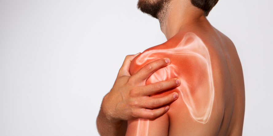
A proximal humerus fracture (or humeral head fracture) is a type of fracture affecting the shoulder area. This injury occurs in the bone of the upper arm near the glenohumeral joint, which is what most people think of as the shoulder joint.
The humeral head is the rounded top of the humerus (upper arm bone) that forms the “ball” in the ball-and-socket structure of the shoulder that fits into the shoulder socket (the glenoid cavity of the scapula) forming the shoulder joint and allowing for a complex and dynamic range of motion. Fractures in this area can be challenging to treat due to the complex anatomy and the involvement of crucial structures like the rotator cuff. The rotator cuff muscles and tendons play a vital role in shoulder stability and movement, and injuries to this area can significantly impact function.
A proximal humerus fracture can significantly impact overall function and stability of the shoulder, causing pain, limited range of motion and potential complications like arthritis or joint stiffness, if not treated properly. These fractures can also cause long-term impairments, including residual pain and strength deficits.
A proximal humerus fracture, like many others, may be caused by various factors such as a trauma or a fall, a sports injury or osteoporosis. One of the main causes is trauma, such as a direct blow to the shoulder (e.g., a fall, a collision in sports or physical assault) or falling onto an outstretched arm (a common mechanism in both young and elderly people).
These fractures are commonly seen in individuals involved in contact sports (e.g., football) or as a result of motor vehicle accidents.
Another significant factor is osteoporosis. It is a degenerative bone condition where bones become porous and fragile and it is more common in postmenopausal women and elderly people. With age, bone strength decreases, making the humeral head more susceptible to fracture even with minimal force. These fractures may occur from low-impact events such as tripping and falling at home or lifting something heavy awkwardly. In elderly people, such injuries are often associated with more complex fracture types. These may involve displacement, where bone fragments shift from their normal alignment (position).
When a proximal humerus fracture occurs, symptoms vary depending on severity. The severity of symptoms depends on the nature and the extent of the fracture (e.g., whether it is displaced or not). The most common signs include:
Additionally, when the fracture is displaced (the broken bone fragments are out of alignment) there is a higher risk of nerve and blood vessel damage. The axillary nerve is the most commonly injured in such cases, leading to numbness over the outer shoulder and weakness in lifting the arm. Although less common than nerve injury, blood vessels near the shoulder joint (such as branches of the axillary artery) can also be compromised, especially with high-energy trauma or displaced fractures. This can lead to poor circulation, increased swelling, or even compartment syndrome (a rare but serious condition).
The diagnostic process for a proximal humerus fracture begins with a thorough clinical examination, which includes: history taking, inspection and palpation range of motion testing and neurovascular assessment. Once a fracture is suspected, imaging tests are essential to confirm the diagnosis and plan treatment. These tests include X-rays that helps determine if the fracture is non-displaced, displaced, intra-articular, or if there are associated injuries like dislocations or fractures in other parts of the shoulder joint. CT Scan is especially useful for comminuted fractures (where the bone is broken into several pieces) or those that involve the joint surface. The imaging tests are critical in pre-surgical planning, helping surgeons assess the bone alignment and evaluate the extent of fragmentation. This detailed information enables them to plan the surgical procedure more accurately, select the most appropriate surgical technique, and predict potential challenges.
A proximal humerus fracture refers to a break in the upper part of the humerus (the arm bone that fits into the shoulder socket). Treatment varies depending on several factors, including the fracture type and extent, the patient’s age, overall health and activity level.
The stable, non-displaced fractures can often be treated conservatively. This includes immobilizing the arm with a sling to allow natural healing, pain management, and starting physiotherapy to manage discomfort and restore the range of motion and strength in the shoulder. The sling is typically removed after 6 weeks. This method avoids surgery and is preferred in older or low-demand patients, or where the fracture is expected to heal well on its own.
When the fracture is unstable or displaced (bone pieces have shifted or are no longer aligned), surgery is usually necessary to realign and stabilize the bone to ensure proper healing and restore function.
The appropriate surgical approach is selected after evaluating a number of factors, such as bone quality, fracture orientation and any potential accompanying soft tissue injuries.
Key factors during pre-surgical evaluation include patient age, overall health, and desired activity level. The surgery of choice is open reduction and internal fixation (ORIF) where bone fragments are surgically repositioned and held together with plates, screws, or pins. This method is typically used in younger or active patients with good bone quality.
In severe cases with comminuted fractures (many fragments) or when the blood supply to the humeral head is at risk (risk of avascular necrosis), the damaged bone is replaced with a metal implant. This is common in elderly patients or those with poor bone quality.
Physiotherapy plays a vital role in both surgical and non-surgical cases. It helps patients recover from injuries or surgeries by focusing on regaining muscle strength, flexibility, and joint mobility. Each patient’s rehabilitation program is specifically designed based on their injury severity, type of treatment (conservative vs. surgical), and personal needs. Conservative treatment may involve rest, immobilization, and gradual movement, while surgical treatment requires post-op physiotherapy to restore function.
to ensure recovery—by restoring muscle strength, flexibility, and joint range of motion. Rehabilitation is tailored to each individual case.
Proximal humerus fractures are complex injuries requiring detailed knowledge of shoulder joint anatomy and surgical expertise. The main goal is to restore shoulder function (mobility and strength), relieve pain and prevent complications and enable patients return to normal daily activities, and if applicable, work or sports.
The Orthopedic Upper Limb Surgeon Dr. Panagiotis Pantos has specialized knowledge and extensive experience in managing numerous shoulder area fractures, always choosing the treatment approach that best suits both the fracture type and patient needs.

Fill in your details below and we will contact you immediately!
WIN THE
MATCH POINT IN THE RACE
OF YOUR HEALTH!